
Located at the Jackson Clinic Baptist Campus, The Women’s Imaging Department centralizes many of the diagnostic services offered by the clinic for women and incorporates the latest technology in testing procedures. Patients can have tests done on digital mammography equipment and ultrasound for early detection of breast cancer, or they can screen for the presence of osteoporosis with digital bone density testing equipment.
The Jackson Clinic is accredited through the American College of Radiology. The Jackson Clinic’s mammography staff is accredited through the American Registry of Radiologic Technologists
Digital Bone Density
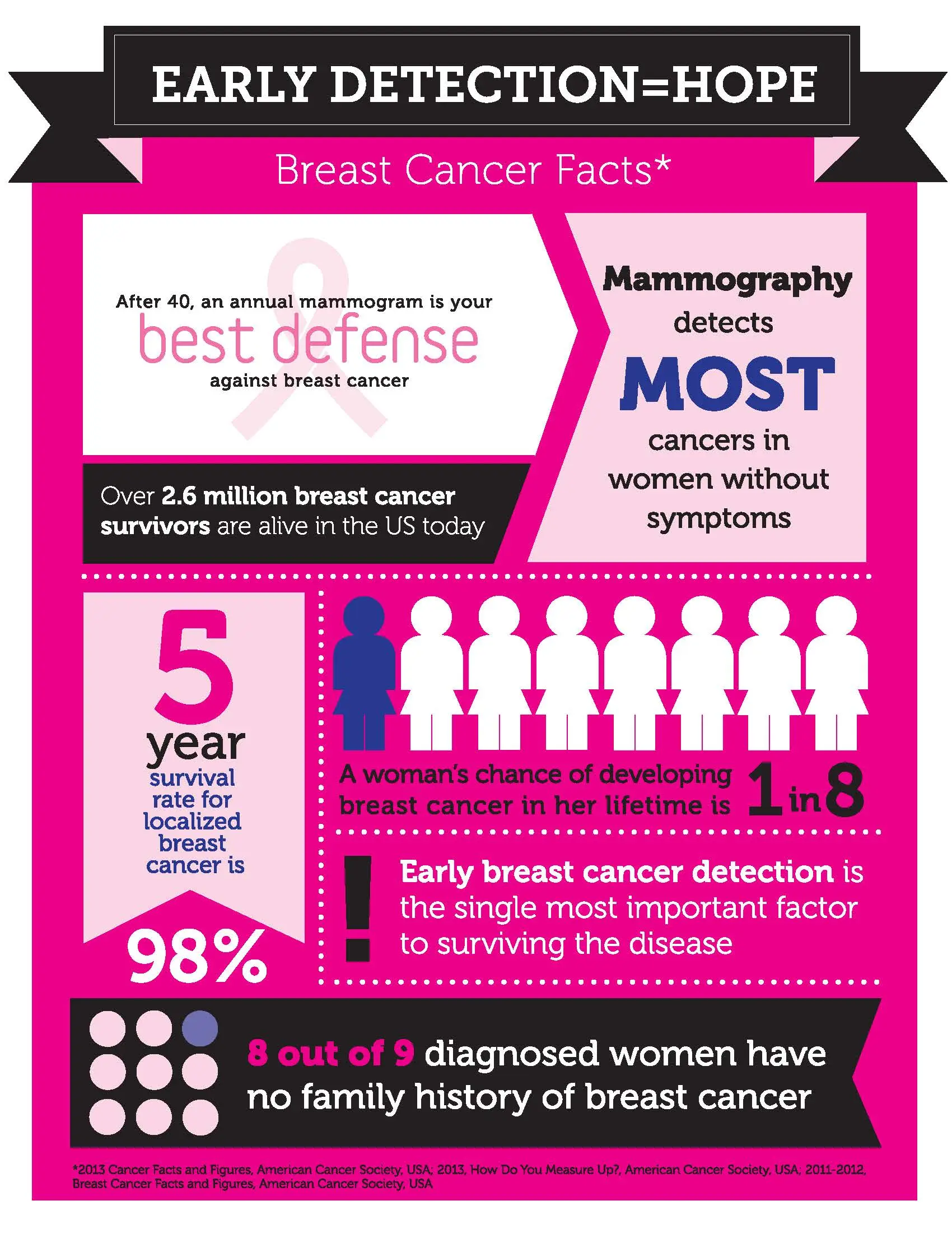
Bone density tests help doctors to diagnose the presence or risk of osteoporosis. Osteoporosis is caused when bones become thin and brittle, which increases the risk of a fracture - many patients do not realize that they have osteoporosis until a fracture occurs. The earlier osteoporosis is diagnosed, the more likely future fractures can be prevented.
Getting a bone density test is easy with our DEXA bone density equipment. DEXA, or Dual Energy X-ray Absorptiometry, uses low energy x-rays to measure the density of bones, muscle and body fat. The procedure is quick, painless and highly accurate. It scans the spine and hips because the bone density there is indicative of bone density elsewhere in the body.
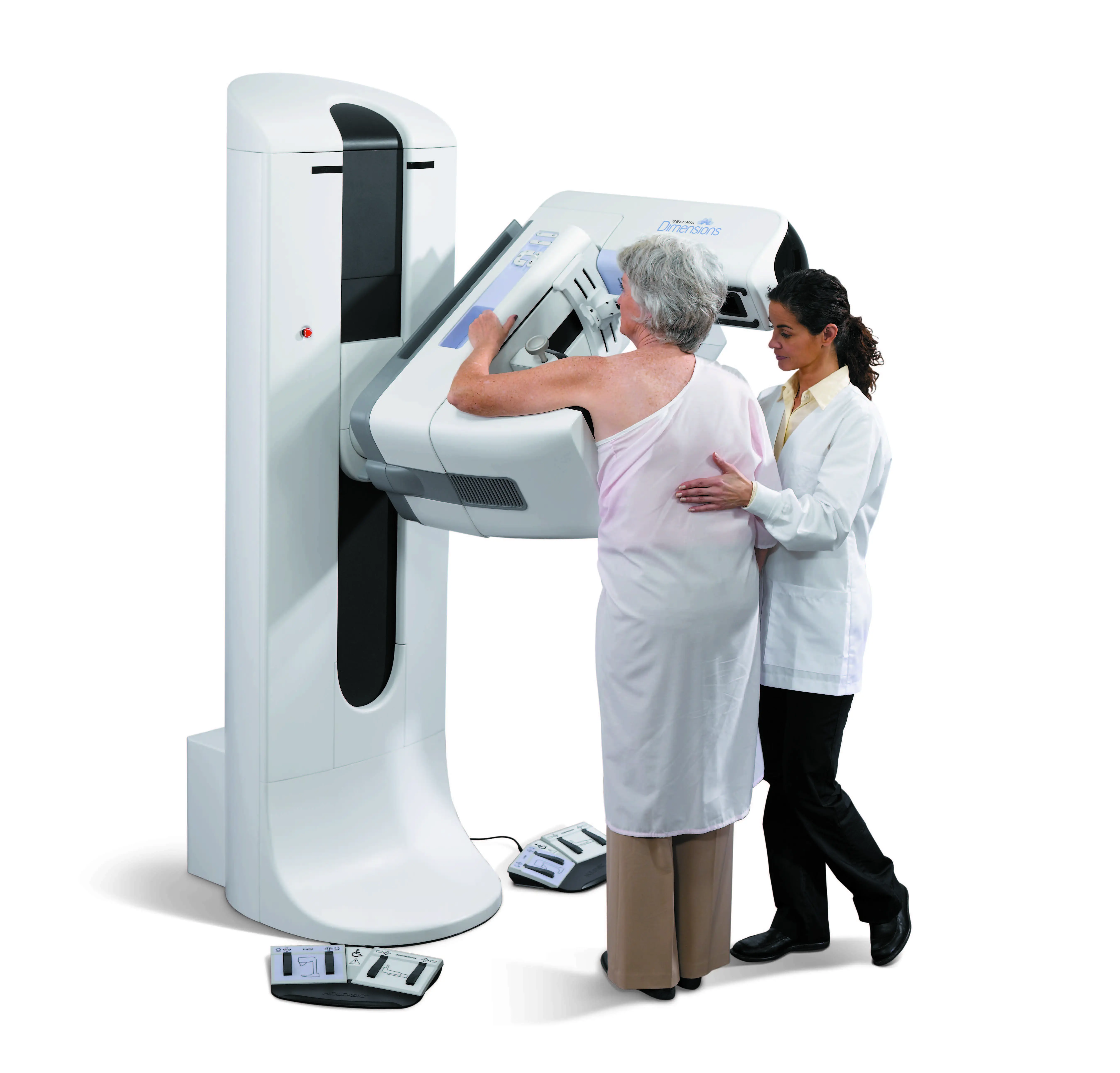
Digital Mammography and Breast Tomosynthesis (3D Mammography)
Digital mammograms take digital images of the breast, allowing doctors to magnify and change the contrast of an image, resulting in improved chances of detecting breast cancer early. The digital technology also allows the mammographers to quickly see the quality of the image and redo it is need be.
What is Breast Tomosynthesis?
Breast tomosynthesis - often called 3D Mammography or digital tomosynthesis - is a breakthrough in mammography that provides a clearer, more accurate view compared to traditional mammography alone.
It is similar to a traditional digital or 2D mammogram with the same level of radiation and breast compression, but detects 41% more invasive breast cancers and reduces false positives by up to 40% and only takes a few seconds longer. The Jackson Clinic Women’s Imaging Department uses the Hologic Genius 3D Mammography technology approved by the FDA.
A screening experience is similar to a traditional mammogram. During the mammography exam, multiple, low-dose images of the breast are acquired at different angles. These images are then used to produce a series of one-millimeter thick slices that can be viewed as a 3D reconstruction of the breast. A three-dimensional view of the breast tissue is produced that helps radiologists identify and characterize individual breast structures without the confusion of overlapping tissue. It allows doctors to see masses and distortions associated with cancer and precancerous cells significantly more clearly than conventional 2D mammography.
Our 3D mammography benefits all screening and diagnostic mammography patients, especially women receiving a baseline screening, who have dense breast tissue, and/or have a personal history of breast cancer.
The benefits of Breast Tomosynthesis include:
- 41% increase in the detection of invasive breast cancers
- 29% increase in the detection of all breast cancers
- 15% decrease in women recalled for additional imaging
- 49% increase in Positive Predictive Value (PPV) for a recall
- 21% increase in PPV for biopsy.